DR.CHETANA(INTERN)
DR.ABDUL RAHEEM (INTERN)
DR.ASHFAQ(INTERN)
DR.SRAVYA(INTERN)
DR.GNANADA(INTERN)
DR.CHARAN(PG1)
DR.VAMSI(PG1)
DR.SUSMITHA(PG2)
DR.ADITHYA (PG3)
DR.PRANEETH(PG3)
DR.PRAVEEN NAIK (ASS.PROF)
DR.RAKESH BISWAS(HOD)
This is an online E log book to discuss our patient's de-identified health data shared after taking his/her/guardian's signed informed consent.
Here we discuss our individual patient's problems through series of inputs from available global online community of experts with an aim to solve those patient's clinical problems with collective current best evidence based inputs.
This E log book also reflects my patient-centered online learning portfolio and your valuable inputs on the comment box is welcome.
Here is a case i have seen:
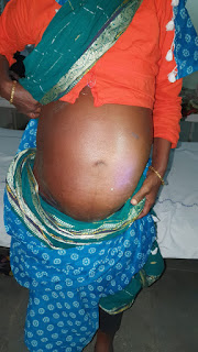
A 45y/o female, from West Bengal, presented to casualty with complaints of abdominal distention since 2 years & pedal edema since 2 months.
Patient was apparently asymptomatic 2 years back.Then developed abdominal distension , insidious in onset and has been rapidly progressing since last one year.
With the increased distention she complains of SOB, difficulty to eat and drink and extreme low back ache.She has desire to eat varieties but is unable to.On doing paracentesis there is improved urine output and decreased SOB ,increased apetite.
Pedal edema since 2 months which is more in the mornings.
She’s k/c/o DM since one year.Took medicines for 6 months and stopped.
Not a k/c/o HTN , TB, Thyroid or Cardiac diseases.
No significant Family history. Has 7 children ( 4 sons , 3 daughters)
Menopause -4.5 years ago.
Bowel movements irregular, sleep inadequate , No addictions.
No h/o. Drug Allergies
General Examination-
Pt is conscious, coherent and cooperative.
Malnourished.
Clubbing +
? Lymphadenopathy- Localised, around umbilicus.
Terry nails +
No Pallor, Icterus, Cyanosis , Koilonychia, Palmar erythema.
Vitals:-
Temperature- 98.5 F
Bp - 110/90 mm Hg
PR - 64 bpm , regular
RR -24 cycles / min
P/A:
Inspection- Ovoid distension of abdomen , slit like umbilicus, absence of scars and sinuses , engorged veins+
Palpation- ? Lymph nodes palpated around umbilicus -multiple, smooth, regular , adherent, non-tender and immobile.
Hepatojugular reflux sign -nt.
Percussion- Fluid Thrill +nt
Auscultation: BS+
CVS: S1 S2 + , no murmurs
RS: Dull note on percussion & Decreased air entry on right side
CNS: NFND
INVESTIGATIONS:
Ascitic tap:
Ascitic Fluid Cell Count
TC 192
N 60%
L 40
RBC plenty
Pleural tap:
Serology: Negative
X-ray chest:PA
Triple phase CECT
DIAGNOSIS:
ASCITES with PORTAL HTN
RIGHT PLEURAL EFFUSION
Treatment:Day 1 to Day 8
Fluid restriction less than 1litre per day
Salt restriction less than 2 gms per day
Tab.Lasilactone
BP/PR/TEMPERATURE MONITORING 4TH HOURLY
Ascitic tap done on day 1 ,5litres tapped
On analysis it is high SAAG low protien,it is probably due to portal HTN ,
QUESTION:
what is the cause of her portal hypertension?
Pleural tap was done on day 2
On analysis according to lights criteria it transudate.
?.Hepatic hydrothorax
On day 7
Ascitic tap and pleural tap was done at same time sent for analysis:
Right sided:Pleural fliud
Left sided: Ascitic fluid
ASCITIC FLUID. PLEURAL FLUID
Sugars. 109mg/dl. 75mg/dl
Protein. 1.5 gm/dl. 2.1 gm /dl
LDH. 214 IU /L. 234 IU/L
SAAG. 1.5
CELL COUNTS
TC. 21 cells. 25 cells
DC.
lymphocytes. 80 percent. 40 percent
neutrophils 20 percent. 60percent
RBC. present. Plenty
DAY8 _Therapuetic ascitic tap done 2 litres tapped
DAY 9 &DAY 10
Fluid restriction less than 1 litre per day
Salt restriction less than 2 gms per day
TAB.LASIX 80 mg BD
TAB.ALDACTONE 50 mg BD
ON DAY 10 :
24 HRS URINE VOLUME:1,100ML
24 HRS URINARY CREATININE:0.42GM/DAY
24 HRS URINARY SODIUM:278mmol/day
DAY 11 to DAY 14
Fluid restriction less than 1 litre per day
Salt restriction less than 2 gms per day
TAB.LASIX 20 mg BD
TAB.ALDACTONE 50 mg BD
SERUM CREATININE_0.9MG/DL
Day 11: Ascitic tap done 2 litres tapped
Day 14:
MR venogram done:
Shrunken right lobe of liver with hypertrophy of left lobe
Gross ascitis
Gross right sided pleural effusion with pleural thickening and collapse of entire lung
Multiple vertebral bodies and rib lesions in the visualised dorsal and lumbar spine
Severe narrowing of the intrahepatic portion of inferior venacava with cranial most aspect of
showing very faint intraluminal opacification on contrast administration suggestive of chronic budd chiari syndrome
Rule out sarcoidosis and metastasis.
DAY 15:
Fluid restriction less than 1 litre per day
Salt restriction less than 2 gms per day
TAB.LASIX 20 mg BD
TAB.ALDACTONE 50 mg BD
TAB.WARFARIN 5MG OD
INJ.ENOXAPARIN 40 MG OD
Ascitic tap done:2 litres tapped
DAY 16:
Fluid restriction less than 1 litre per day
Salt restriction less than 2 gms per day
TAB.LASIX 40 mg BD
TAB.ALDACTONE 50 mg BD
TAB.WARFARIN 5MG OD
Heparin was withholded
Started prednisolone 30 mg OD
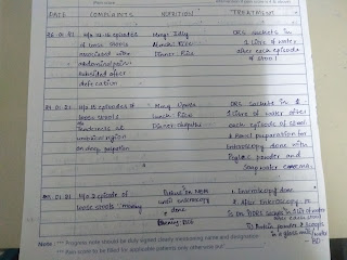
APRAXIA CHARTING WAS DONE:

Day-17-19: planned for iliac crest biopsy
Fluid restriction less than 1 litre per day,
Salt restriction less than 2 gms per day,
TAB.LASIX 40 mg BD,
TAB.ALDACTONE 50 mg BD.
Anticoagulants and steroids are with holded until iliac crest biopsy.
DAY-20:
BONE BIOPSY DONE:CT GUIDED ,FROM POSTERIOR SIDE OF ILIAC CREST UNDER LA TO
RULE OUT THE MULTIPLE HYPERDENSE LESIONS IN THE BONES{SPINE,RIBS,PELVIS};
Fluid restriction less than 1 litre per day,
Salt restriction less than 2 gms per day,
TAB.LASIX 40 mg BD,
TAB.ALDACTONE 50 mg BD.
Day-21:
ORTHOPEADIC REFERRAL DONE FOR RESTRICTRD LEFT SHOULDER MOVEMENTS SINCE 4
MONTHS
ADVISED: X RAY left shoulder : F/s/o asthetic changes
advised:
TAB.DOLONEXT DT BD FOR ONE WEEK,
TAB.PAN 40 MG OD ONE WEEK,
TAB.TENDOFIT OD FOR ONE MONTH,
PHYSIOTHERAPY IFT/TENS LEFT SHOULDER WITH SHOULDER ROM EXERCISES.
Day-22 -23
Fluid restriction less than 1 litre per day,
Salt restriction less than 2 gms per day,
TAB.LASIX 40 mg BD,
TAB.ALDACTONE 50 mg BD,
TAB.WARFARIN 5 MG PO OD
TAB.PREDNISOLONE 30 MG PO OD
TAB.DOLONEXT DT BD FOR ONE WEEK,
TAB.PAN 40 MG OD ONE WEEK,
TAB.TENDOFIT OD FOR ONE MONTH,
PHYSIOTHERAPY IFT/TENS LEFT SHOULDER WITH SHOULDER ROM EXERCISES.
Bone biopsy results:
DISCUSSION:
?.cause of portal hypertension in this pt.
?. cause of refractory ascitis.
https://www.ncbi.nlm.nih.gov/pmc/articles/PMC2886420/
https://www.ncbi.nlm.nih.gov/pmc/articles/PMC2886420/
Cause of pleural effusion ?Hepatic hydrothorax
Most probably because both are transudate on both fluid analysis at same time appears to be same.
What are the complications of ascitis ????
Hepatic hydrothorax
Spontaneous bacterial peritonitis
Any other????
Reason for sclerotic changes in spine,ribs and pelvis?????
Still a doubt!!!!! Sarcoidosis??????
Is focal segmental stenosis of intrahepatic Ivc suggests Budd chiari syndrome????
MR VENOGRAPHY FINDINGS OF OBSTRUCTED HEPATIC VEINS AND HEPATIC VEINS AND THE INFERIOR VENACAVA IN BUDD CHIARI SYNDROME.
"Segmental lesions were identified as obstruction length > 1.0 cm in HVs and > 1.5 cm in the IVC (5,6). Segmental lesions were also classified as segmental obstruction or segmental stenosis. The lumen and blood disappeared on MRI in the presence of segmental occlusion (Fig. 1). Segmental stenosis was seen as a stenosed lumen with a thickened wall."
"Thrombi in HVs, accessory hepatic veins (AHVs), and the IVC were seen as diverse intensities in different MR sequences during the development of thrombi. MRI revealed an absence of blood flow in the vessels that were completely thrombosed by fresh thrombi. Furthermore, where there was partial embolization, there was a blood-filling defect in the vessel"
A case of mltisystemic sarcoidosis presenting as budd chiari syndrome.
"A 42-year old female was referred to our department for chronic anicteric cholestasis discovered fortuitously through a regular blood test. Her past medical history revealed diabetes mellitus evolving for 3 years; she had never received oral contraceptive therapy. She described progressive worsening cough and exertional dyspnea. She denied right upper quadrant pain, fatigue or pruritus. On examination, her liver span was 21 cm and spleen was palpable 4 cm below the left costal margin. Skin examination disclosed red-brown papular plates measuring 2 to 5 mm in diameter and having a predilection on scars and sites of trauma (Figure 1). Cardiovascular and respiratory examination was normal. Liver tests demonstrated increased cholestatic enzymes: a γ-glutamyl transferase level of 136 UI/L (normal: 12-58) and ALP of 325 UI/L (normal: 38-126) without hyperbilirubinemia (total bilirubin 13 mg/L; normal: 2-12); aminostransferases levels were normal. Other laboratory tests revealed normocytic inflammatory anemia with hemoglobin of 10.6 g/dL, mean corpuscular volume of 82.8 fL and serum ferritin of 150 g/L; white blood cell count of 4700/mm3 and platelets count of 221,000/mm3. Erythrocyte sedimentation rate was 85 mm at 1 h. International normalized ratio was normal. Serum and urinary calcium were normal."
"Moreover, celiac, hepatic pedicle and lomboaortic adenopathies were noticed. We completed with a CT scan of the chest, which demonstrated mediastinal and hilar adenopathies with multiple micronodular opacities on the lower pulmonary lobes. At screening upper endoscopy for portal hypertension, there were neither esophageal nor gastric varices but gastric mucosa was erythematous. Gastric biopsies were performed and disclosed non-caseating granuloma. Likewise, liver biopsy showed the presence of sarcoid granulomas consisting of a compact aggregate of large epithelioid cells, sometimes with multinucleated giant cells and a surrounding cuff of lymphocytes (Figure 3). These tend to be more frequent in portal tracts. Caseation or damage bile ducts were not present."
What is gold standard investigation to diagnose Budd chiari syndrome???
MRI OR USG ABDOMEN,In this pt we did MR venogram to detect any intrahepatic ivc obstruction.
14.3.2021_
Unfortunately patient was expired today at home,cause of death is uncertain.


